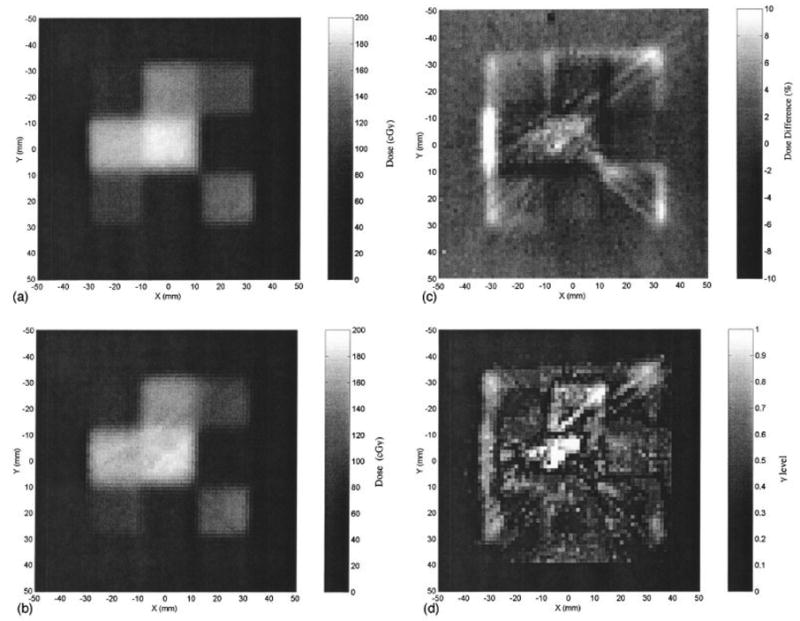
Figure 7.

A radiochromic measurement of a simple IMRT field (a) is compared against an optical-CT image of the same plane (b). The percent dose difference between radiochromic film and gel measured dose-maps is shown in (c), the corresponding gamma maps (1.5mm and 5% dose criteria) in (d). Significant streak and cupping artifacts are visible in the optical images. From Islam et al [10].
