
Figure 2.
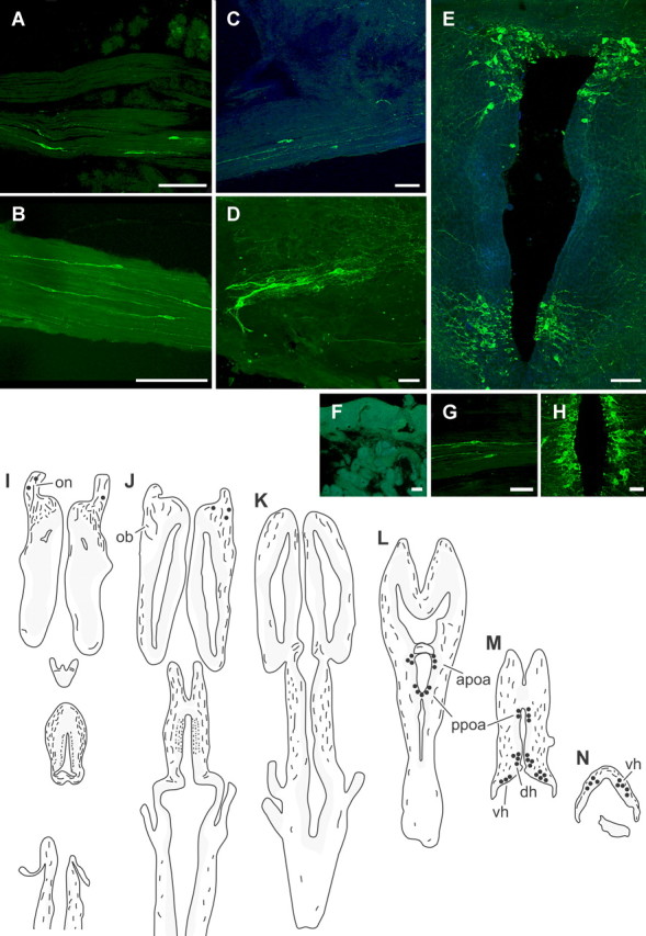
Distribution of NPY-like immunoreactivity in the brain and snout of adult axolotls. A–H, Confocal images of NPY-immunoreactive terminal nerve cell bodies and fibers (green); for clarity, some sections have been counterstained with Hoechst 33258, a fluorescent Nissl stain (blue). Anterior is toward the left in A–D and G, and toward the top in E and H–N; the lumen is toward the top in F. Scale bars, 100 μm. A, Olfactory nerve fascicles underneath the olfactory epithelium containing labeled terminal nerve cells. B, Labeled terminal nerve neurons inside the olfactory nerve. C, A cluster of labeled neurons in the ventral olfactory bulb. D, Labeled cells in the proximal portion of the olfactory nerve, adjacent to the olfactory bulb. E, Montage of three images illustrating labeled cells in the anterior and posterior preoptic area. F, No NPY-like immunoreactivity was observed in the olfactory epithelium. G–H, Images showing the distribution of NPY immunoreactivity in control experiments. G, NPY-immunoreactive terminal nerve cells in olfactory nerve fascicles after preincubation of anti-NPY antiserum with GYIRFamide. H, Labeled cells in the posterior preoptic area after preincubation of anti-NPY antiserum with FMRFamide. I–N, Illustrations showing the distribution of labeled cells and fibers in horizontal sections [from dorsal (I) to ventral (N)] through the brain of an adult axolotl. Shaded areas represent locations of large groups of neurons. Filled circles indicate regions in which labeled neurons were observed. apoa, Anterior preoptic area; dh, dorsal hypothalamus; ob, olfactory bulb; on, olfactory nerve; ppoa, posterior preoptic area; vh, ventral hypothalamus.