
Fig. 1. TGFβ1 reverses the decline in vimentin gene expression during C2C12 differentiation.
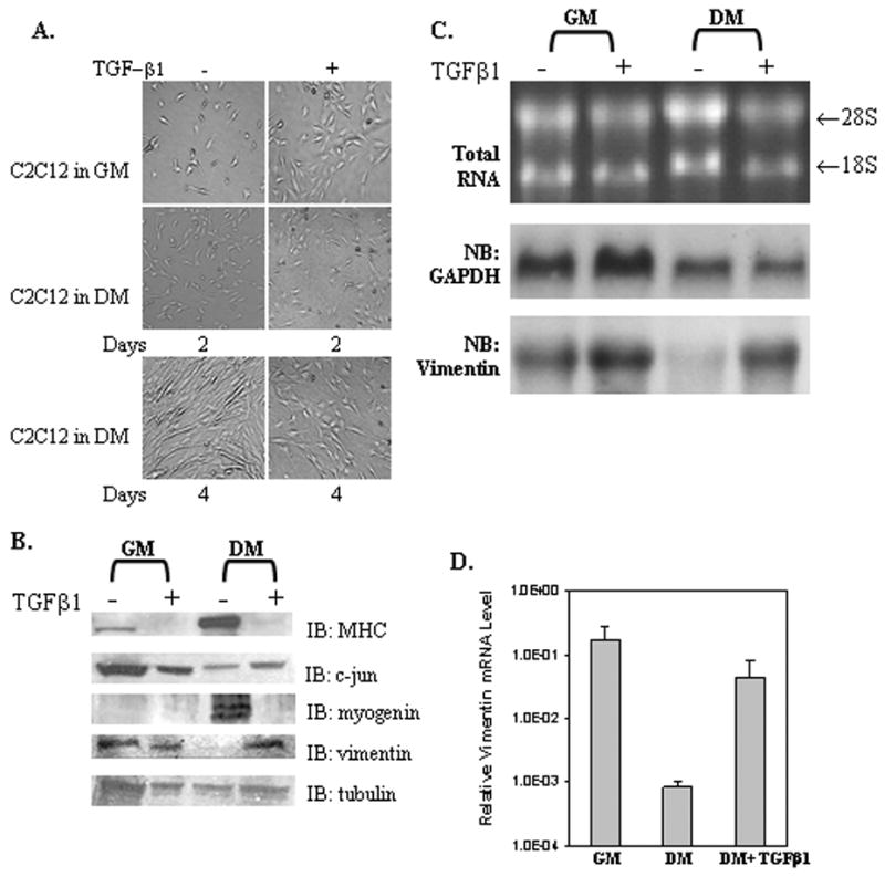
(A) C2C12 cells grown in DMEM medium plus 15% FBS (growth medium, GM) or 2% horse serum (differentiation medium, DM) without or with TGFβ1 (1 ng/ml) for 2 or 4 days. (B) Western blot analysis of WCEs (50 ug) isolated from C2C12 cells as grown in panel A for 4 days and described in Materials and Methods. (C) Northern blot analysis of total RNA (20 μg) isolated from C2C12 cells grown in GM or DM minus or plus TGFβ1 for 4 days as previously described [32]. The nitrocellulose membrane was first hybridized with a 32P-labeled human vimentin cDNA probe (bottom) and then the membrane was stripped and re-probed with a 32P-labeled GADPH cDNA (middle). Another sample of total RNA (5 μg) was stained with ethidium bromide to verify the integrity of the RNA (Top). (D) Total RNA (100 ng) was reverse-transcribed into cDNA using poly-dT as a primer followed by the use of 1/20 of this sample for real-time PCR analysis. The data was normalized to adolase gene expression. The Y-axis represents the relative mRNA levels of the vimentin gene. Results are the average of three separate experiments performed in triplicate with bars representing the standard error.