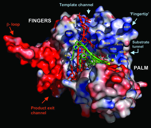
Fig. 4.
RNA modeling onto IBDV VP1. The dsRNA duplex is brought from reovirus elongation complex by superimposing 61 conserved Cα atoms, as described in Table 1. The VP1 model in this figure contains residues 169–579. The thumb, N-terminal, and C-terminal domains have been removed from the molecule to achieve a better view of the polymerase active site. Important amino acid residues are labeled. Molecular surface is colored according to electrostatic potential. In the active site, the template is shown in red; nascent, green; metal ion, yellow spheres; nucleotide substrate, pale blue.

