
Fig. 2. Activation of the c-FLIP Promoter by AR.
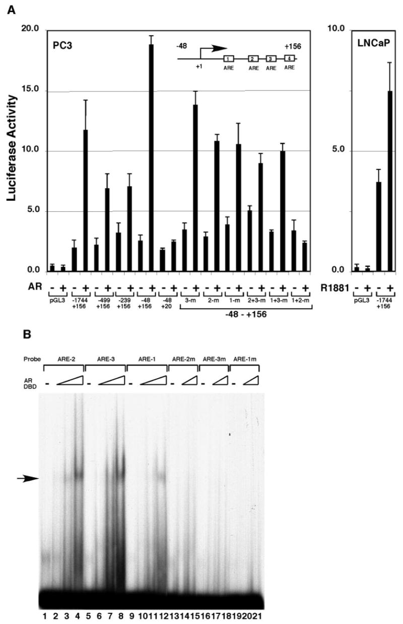
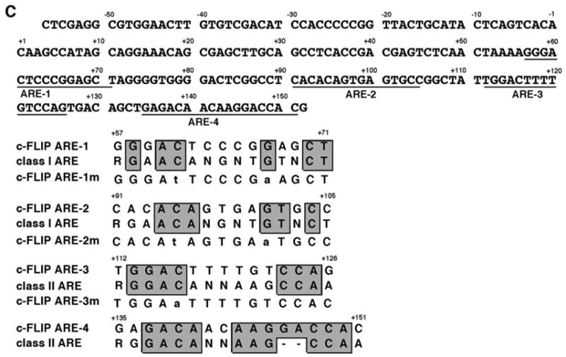
A, PC3 cells were transfected with 100 ng of the reporter plasmids containing various truncations and point mutations of the c-FLIP promoter and with or without 150 ng pcDNA-AR. LNCaP cells were transfected with 100 ng of the reporter plasmid containing the c-FLIP promoter (−1744 to +156) and grown in the absence or presence of 10 nM R1881. B, Binding of c-FLIP AREs to the DBD of AR. The different 32P-labeled ARE sites in the c-FLIP promoter were incubated with 0 ng (lanes 1, 5, 9, 13, 16, and 19), 20 ng (lanes 2, 6, and 10), 60 ng (lanes 3, 7, 11, 14, 17, and 20), or 180 ng (lanes 4, 8, 12, 15, 18, and 21) ng of the recombinant DBD of AR. The complexes of AR-DBD-ARE are indicated by an arrow at the left. C, The nucleotide sequence of the ARE-containing region in the c-FLIP promoter and the alignment of the c-FLIP AREs with the consensus AR binding sequences. The point mutations used in transient transfection and gel shift assays are indicated in lowercase letters.