
Figure 1.
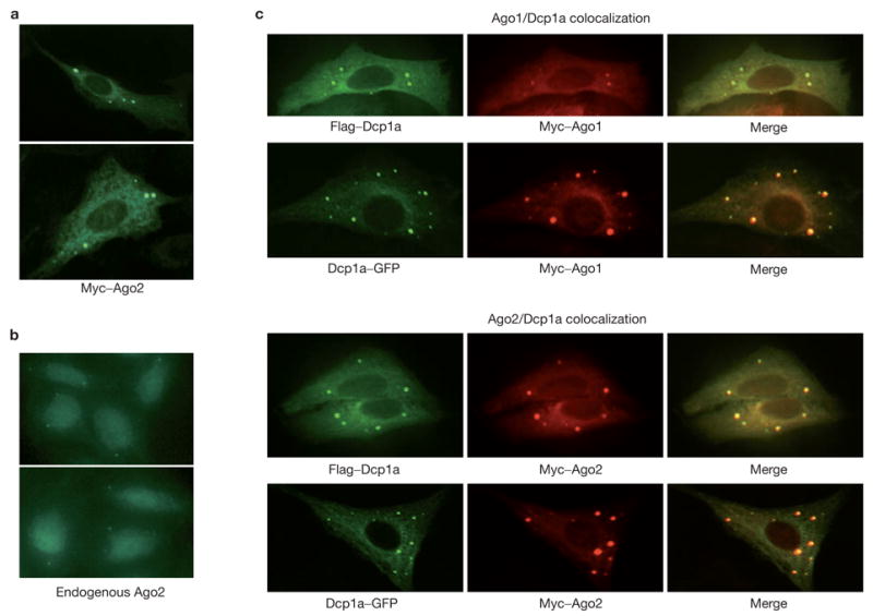
Argonaute proteins localize to mammalian P-bodies. (a) Myc-tagged Ago2 protein was expressed in U2-OS cells. Ago2 protein localized to discrete cytoplasmic foci by staining with FITC-conjugated anti-Myc. (b) Endogenous Ago2 protein was localized in U2-OS cells by staining with a rabbit anti-Ago2 antibody. (c) Argonaute proteins colocalized with either GFP- or Flag-tagged Dcp1a, a signature component of the mammalian P-bodies. Argonaute proteins were visualized using a Rhodamine-Red-conjugated anti-Myc. Dcp1a was visualized either by GFP or FITC-conjugated anti-Flag.