
Figure 1.
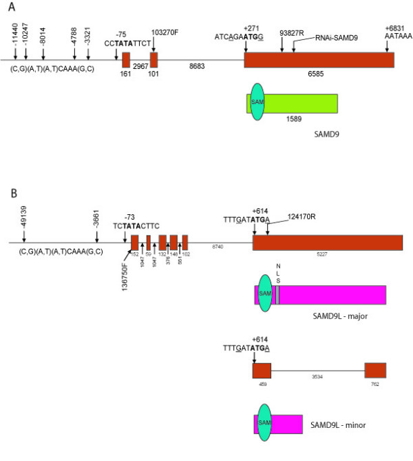
Gene and protein structure of SAMD9 and SAMD9L. A. Diagram of SAMD9 gene structure and predicted protein structure. Exons are shown as red rectangles. The protein is shown in green, with the sterile alpha motif domain as blue oval. B. Diagram of SAMD9L gene structure and predicted protein structure. Exons are shown in red. The protein is shown as a pink box, with two predicted open reading frames of shown. The sterile alpha motif domain is shown as blue oval. A potential nuclear localization domain is shown as grey box.