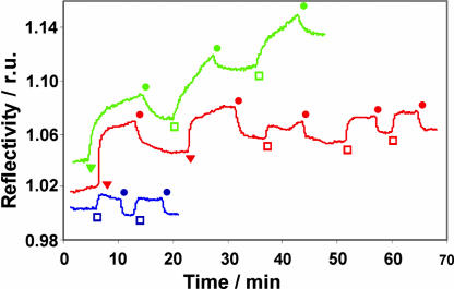
Fig. 7.
SPR sensogram of vesicles of CD1 on βCD SAMs. Blue, vesicles of CD1 on βCD SAMs in the absence of metal(II) and L; red, vesicles of CD1 on βCD SAMs in the presence of Cu(II) and L; green, vesicles of CD1 on βCD SAMs in the presence of Ni(II) and L. Injections: □, 10 μM CD1 vesicles; ▾, 0.1 mM CuL2 or 10 μM NiL3; ●, 1 mM NaHCO3 buffer (pH 9). r.u., relative units.

