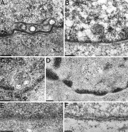
Fig. 5.
Immunoelectron microscopy. Ultrathin sections of RK13-UL31/34 cells. (A) were incubated with rabbit anti-UL34 serum (B) or rabbit anti-UL31 serum (C) or simultaneously with murine anti-UL34 serum and rabbit anti-UL31 serum (D). Bound antibodies were visualized with gold-conjugated secondary anti-rabbit sera containing 10-nm gold for B and C and 15-nm gold for anti-rabbit and 10-nm gold for anti-mouse sera for D. For control, RK13-UL31 cells were labeled with the anti-UL31 serum (E) and RK13-UL34 cells with the anti-UL34 serum (F). (Scale bars, 150 nm.)

