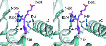
Fig. 3.
Conformational changes of P. falciparum aldolase upon TRAP-tail binding. Shown is a stereoview of the TRAP-binding region. The TRAP-bound structure is shown in light blue, the unliganded structure in gray (29), and the TRAP-tail C-terminal tripeptide DWN in magenta. Key residues binding the penultimate Trp-indole ring are highlighted with sticks. Note the rigid body motion of helix α2, which is part of the helix–loop–helix T42-D66 subdomain shift upon TRAP-binding (see The Indole Anchor-Binding Pocket).

