Fig. 3.
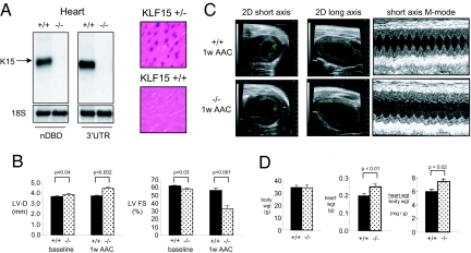
Effect of KLF15 deficiency on cardiac gene expression and pressure-overload hypertrophy. (A) Northern analysis (Left) of wild type and KLF15-null hearts using a cDNA probe coresponding to the non-DNA binding domain (nDBD) or the 3′ UTR of KLF15. β-galactosidase staining of ventricular sections from KLF15 +/+ and +/− mice indicates expression of endogenous KLF15 in cardiomyocytes (Right). (B) KLF15 −/− mice exhibit increased LV dimension and reduced LV systolic function at baseline and with pressure-overload. KLF15 +/+ and −/− mice were subject to AAC and LV diastolic dimension and fractional shortening assessed at 1 week after surgery. A marked reduction in LV function of KLF15 −/− mice is noted after AAC. (C) Representative 2D and M-mode echocardiograms from KLF15 +/+ and −/− animals that were used for the measurements in B and SI Table 1. (D) KLF15 −/− mice hearts (atria and ventricles) after AAC (1 week) are heavier.

