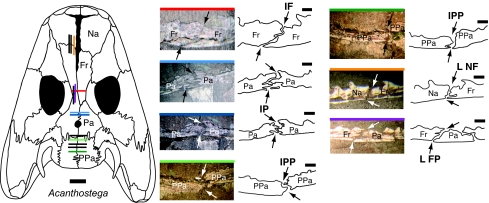
Fig. 2.
Photographs and camera lucida drawings of selected midline and coronal sutures in the aquatic Devonian tetrapod Acanthostega. The midline IF, IP, and IPP sutures were observed in coronal sections of MGUH f.n. 236. The NF and FP sutures were observed in MGUH f.n. 1305 (sectioned sagittally). The black arrows indicate the endocranial and ectocranial emergence of each suture. The colored bar on the top of each suture photograph indicates the approximate position of that slice through the skull roof of Acanthostega. Slices whose positions are shown in black on the dorsal reconstruction of Acanthostega were measured in this study but are not figured here. Note the dramatic shape changes within the IP and IPP sutures. [Scale bars: 1 mm (suture) and 1 cm (skull).] The dorsal reconstruction of the skull of Acanthostega is modified from the literature (31). Na, nasal; Fr, frontal; Pa, parietal; PPa, postparietal; L NF, left nasofrontal suture; L FP, left frontoparietal suture.

