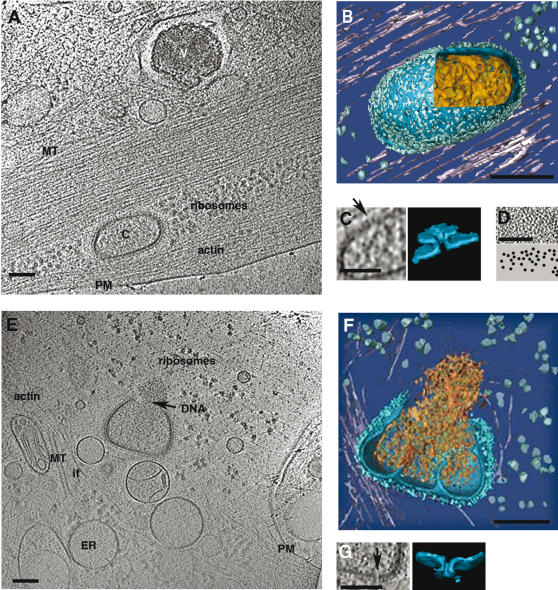
Figure 4. The fate of intra-cellular cores.
PTK2 cells were grown and infected as described under Figure 3 and vitrified at 60 min post-infection. A, a 30 nm thick section through the tomogram (see Movie S2) showing a virion (V) attached to the extracellular side of the plasma membrane as in Figure 3A; and a core (C) in the cytoplasm. After internalization all of the core-features (including arrangement of spikes and DNA) are similar to particles seen before internalization. Actin stress fibers and microtubules (MT) are in the vicinity of the core, (PM–plasma membrane). B, surface rendered core seen in A. C, the pores (arrow) in the core membrane (section and surface rendering). D, a section through the face view of the viral core reveals the random distribution of the spikes in the palisade layer. E and F, a section (30 nm thick) (see Movie S3A) and surface rendered views (see Movie S3B) of the cytoplasm 60 min. after infection. The VV core is opened on one side and releases the genome (arrow, DNA) as an entity. Around the core a number of cellular structures can be observed (PM-plasma membrane, MT-microtubules, ER-endoplasmic reticulum, if-intermediate filaments). G, cross-section and surface rendered views through the core showing one of the 7 nm pores. Bars-100 nm

