Fig. 3.

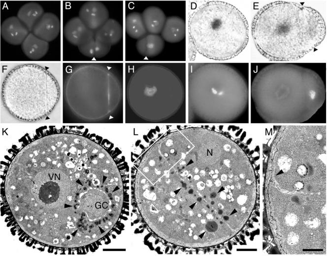
A-C Division axis, cell fate and ultrastructure of gem2 pollen. The division axis was analysed in DAPI stained tetrads of +/gem2; qrt1/qrt1. A, mature qrt1/qrt1 tetrad. B, C, +/gem2; qrt1/qrt1 tetrad showing the conserved plane of division orientated along the polar axis. Arrowheads indicate orientation of the internal wall. D, E, I, J, cell fate analysis in wild-type (D, I) and gem2 showing unequal division (Arrowheads in E, J). Pollen expressing lat52-gus/nia (D, E) and corresponding DAPI images (I, J). F-H, light (F) and fluorescence (G,H) images of a divided gem2 pollen grain showing aniline blue staining of callosic dividing wall (G) and corresponding DAPI image (H). K-M, ultrastructural analysis of wild-type pollen (K) the early bicellular stage. Vegetative nucleus (VN) is located at centrally and numerous lipid bodies (arrowheads) accumulate only in the VC cytoplasm around the generative cell (GC). L, divided gem2 pollen with an internal wall. Lipid bodies (arrowheads) are distributed in both daughter cells adjacent to the internal wall. M, magnified image of junction (arrowhead) region between the dividing wall and the intine shown in L (boxed area). Scale bars, K and L = 3μm, M = 0.5μm
