Fig. 4.

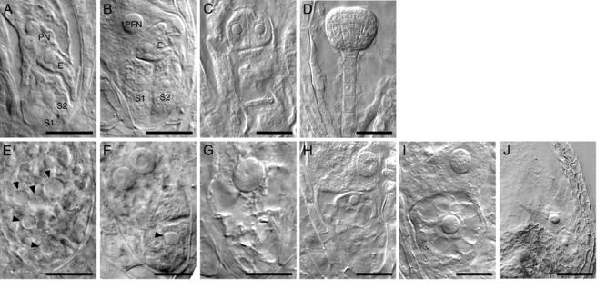
Megagametophyte development in gem2. Nomarski images of wild-type (A-D) and gem2 (E-J) at comparable stages. A, E, eight-cell stage. B, F, G, terminal stage. C, H, I, four-celled embryo stage. D, I, J, late globular embryo stage. A, Wild-type embryo sac with unfused polar nuclei (PN), egg cell (E) and two synergid cells (S1 and S2). B Polar nucleus fused to form large diploid polar fusion nucleus (PFN). C, wild-type embryo showing four-celled embryo proper. D, wild-type globular embryo. E, mutant embryo sacs of gem2 are uncellularized, but contained five nuclei, with various size of nucleoli at the at the micropylar end. F-G Terminal phenotype of a gem2 female gametophyte. Occasionally a mutant embryo sac exhibited a partial wall (F) or a single larger nucleus (G) which is likely to be caused by aberrant nuclear fusion. H-J Mutant female gametophyte of gem2 is unable to form embryo and endosperm as a result of failure of fertilization. gem2 ovule usually contain two nuclei at the micropylar end that is desiccated eventually. Arrowheads (E-J) indicate mutant nucleoli with large size. Scale bars: A-C, E-I = 20μm, D and J = 50μm
