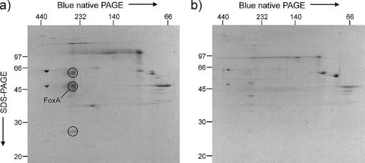
FIG. 5.
Coomassie blue-stained 2D blue native/SDS-PAGE gels of membrane preparations of Sulfolobus metallicus cells grown on pyrite (a) and sulfur (b). The putative FoxA is indicated as one of three spots (circled) that represent a possible protein complex present in pyrite-grown cells but not sulfur-grown cells. The positions of molecular mass markers (in kDa) are shown on the borders of the gels.

