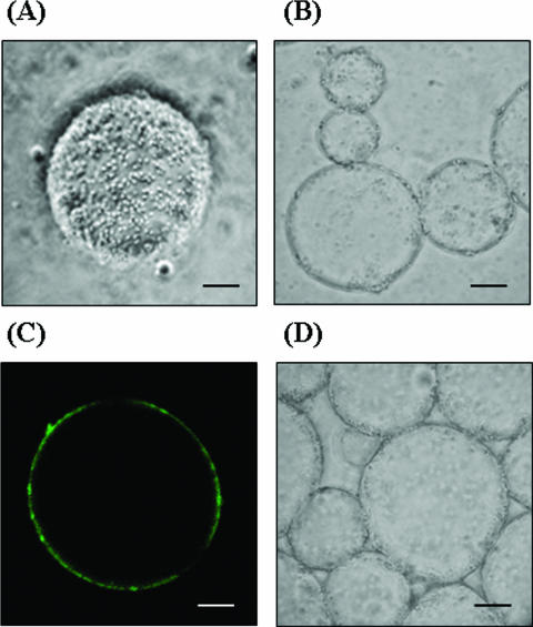
FIG. 6.
Determining the localization of spores at the interfacial surfaces of microemulsions by observing them with various microscopes. Scale bars = 10 μm. (A) Light microscopy of spore of B. subtilis WB700 displaying β-Gal adhering on the surface of microemulsions. (B) Light microscopy of spores of B. subtilis WB700 displaying β-Gal localized at the interface of the water and solvent phases. (C) Confocal fluorescence microscopy of microemulsion of fluorescently labeled spore of B. subtilis WB700 displaying β-Gal using primary rabbit anti-β-Gal antibodies and secondary fluorescein isothiocyanate-labeled anti-rabbit immunoglobulin G antibodies. (D) Light microscopy of control spores of B. subtilis WB700 localized at the interface of the water and solvent phases.

