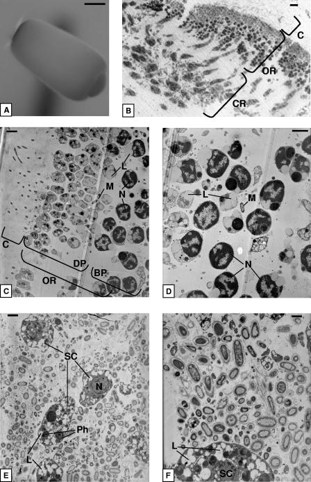
FIG. 2.
(A) Dissection microscopy of a parenchymella larva of I. felix. (B) Light microscopy of a larva cross section showing the ciliary, outer, and center regions. (C and D) Transmission electron microscopy of the ciliary and outer regions showing the distal and the basal parts of sponge cells. (E and F) Transmission electron microscopy of the central part of the larva with electron-transparent sponge cells surrounded by microorganisms. M, microorganisms; BP, basal part; C, ciliary region; CR, central region; DP, distal part; L, lipids; N, nucleus; OR, outer region; Ph, phagosome; SC, sponge cell. Scale bar, 100 μm (A), 10 μm (B), 2 μm (C, D, and E), and 1 μm (F).

