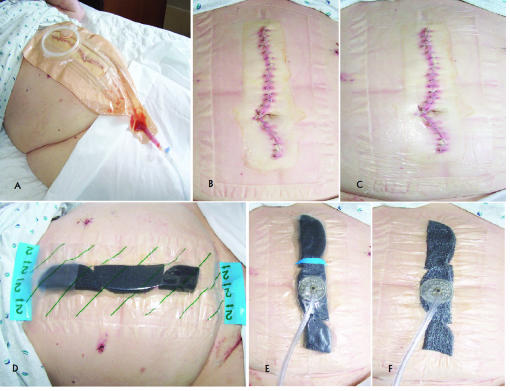
Figure 3.
Stepwise depiction of VAC dressing placement in Case 3. (A) Wound manager overlying the midline wound prior to removal. (B) Midline wound following removal of wound manager and prior to VAC placement. (C) The bottom layer of biocclusive placed on the skin around the incision, with the incision itself left uncovered by biocclusive. (D) VAC sponge placed over the midline incision, along with the second biocclusive (indicated by green diagonal lines). (E) VAC suction device placed over biocclusive prior to application of subatmospheric pressure therapy. (F) The VAC dressing after institution of subatmospheric pressure therapy.

