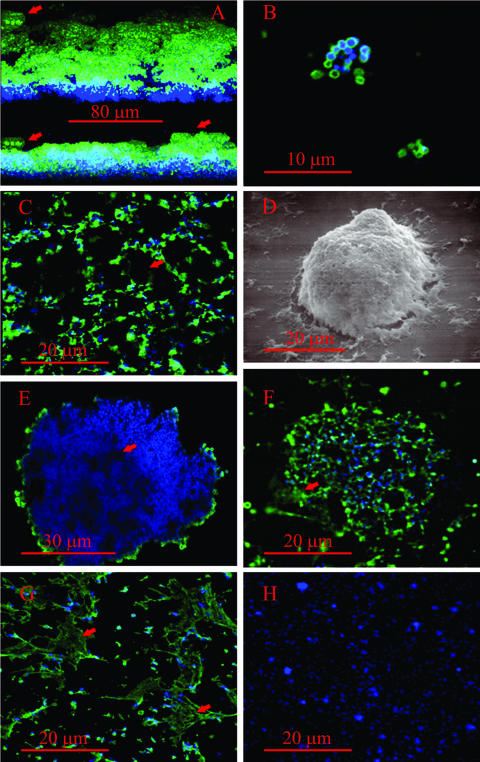
FIG. 2.
Immunofluorescent images of biofilms and microcolonies of M. pulmonis. VsaA epitopes are in green and DNA (Hoechst 33342) is in blue. The scale bars are in red. (A) Three-dimensional reconstruction of a biofilm formed by strain CT183-R3 after 3 days of growth on glass coverslips. The upper image shows the biofilm observed from an angle of elevation of 25 degrees, while the lower image is a horizontal reconstruction of the biofilm. The red arrows point to the tower structures. (B) Laser scanning confocal microscopy image of a small cluster of biofilm-forming mycoplasma cells grown overnight on glass coverslips. (C) Digital image of the honeycombed region. The red arrow points to the cavities in the honeycombs. (D) Scanning electron microscopic image of a tower structure. (E) Cross-sectional image of a biofilm tower acquired by laser scanning confocal microscopy. The arrow points to a channel within the tower. (F) Microcolony of a strain that produces VsaA-R40. The arrow points to extracellular VsaA epitopes. (G) Laser scanning confocal microscopy image of a region of cells adjacent to a microcolony. The red arrow denotes the web-like structures that contain VsaA epitopes. (H) Immunofluorescent image of a biofilm of mycoplasmas that produce the short form of VsaG (control showing that antibody is specific for VsaA).

