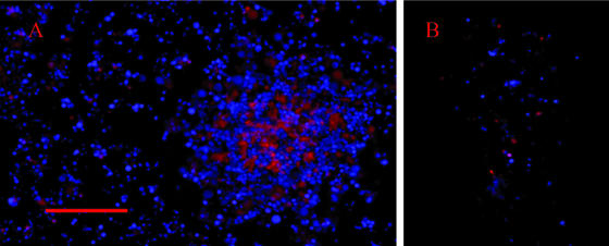
FIG. 3.
DNA staining of a viable (nonfixed) biofilm (A) and a microcolony (B) with Hoechst 33342 and propidium iodide. The biofilms and microcolonies were from cultures that were grown for 2 days. Panel A shows both the honeycombed region of the biofilm and a tower structure (on the right side of panel A). Hoechst staining is shown in blue, and propidium iodide staining is shown in red. The scale bar represents 10 μm.

