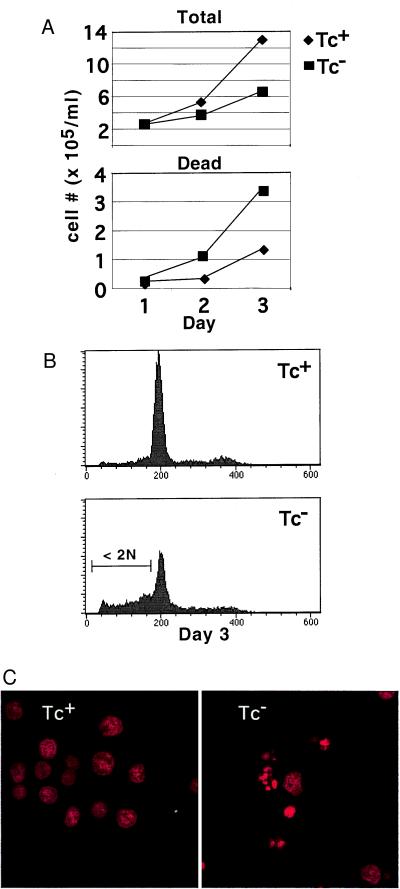
Figure 3.
NF-κB inhibition caused apoptosis. (A) Cell growth was determined by counting on a hemacytometer with the inclusion of trypan blue. (Top) Total cell number—positive and negative for trypan blue. (Bottom) Cell number positive for trypan blue uptake. ⧫, Tc+ media; ■, Tc− media. Cultures were initially seeded at 105/ml and counted at 24-h intervals thereafter. (B) Examination of DNA content on day 3 showed the accumulation of hypodiploid (<2 N) cells. After ethanol fixation, cells were stained with propidium iodide and examined by FACS. (Top) Tc+ media. (Bottom) Tc− media. (C) Confocal microscopy showing apoptotic bodies of cells in B. (Left) Tc+. (Right) Tc−.

