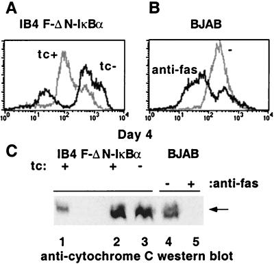
Figure 7.
NF-κB inhibition caused a loss of mitochondrial potential but not Cyt c release. (A and B) DiOC6 fluorescence. (A) IB4 F-ΔN-IκBα cells grown in Tc+ media, gray line, in Tc− media, black line. (B) BJAB, gray line, and BJAB treated with anti-Fas antibody, black line. (C) Cyt c release from the mitochondria. Western blots of S-100 pellets were analyzed for Cyt c. Lane 1, total cell extracts; lane 2, IB4 F-ΔN-IκBα cells, Tc+ media; lane 3, IB4 F-ΔN-IκBα cells, Tc− media; lane 4, BJAB; lane 5, BJAB treated with anti-Fas.

