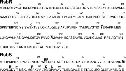
FIG. 3.
RsbR and RsbS mutations. The amino acid sequences of RsbR and RsbS are illustrated. Sites of RsbR and RsbS phosphorylation (i.e., T171 and T205 of RsbR and S59 of RsbS) are in boldface type. Amino acid changes in RsbR and RsbS that were mapped and indicated in Fig. 2 are placed above the original residues in the sequence.

