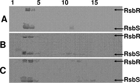
FIG. 6.
Gel filtration chromatography of Rsb proteins in E. coli extracts. Crude extracts were prepared from E. coli strains carrying plasmids pARE7 (PA rsbR rsbS rsbT rsbU) (A), pARE34 [PA rsbR(136EK) rsbS rsbT rsbU] (B), or pARE31 [PA rsbR(225CY) rsbS rsbT rsbU] (C) were fractionated through Sephacryl S-300. Samples from each fraction were analyzed by SDS-PAGE and Western blotting using monoclonal antibodies specific for RsbR and RsbS. Numbers at the top of the figure are fraction numbers, with fraction 1 being the earliest-eluting (high-molecular-mass) fraction. Coomassie-stained gels (not shown) indicate elution of ribosomes between fractions 1 and 5. The positions of RsbR and RsbS on the Western blots are indicated.

