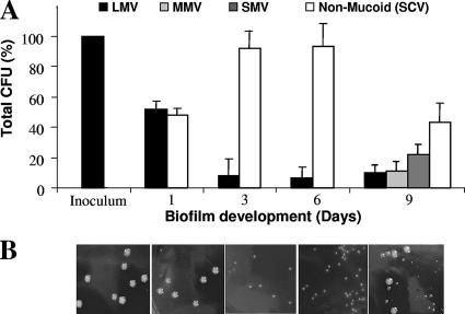
FIG. 2.
Emergence of colony morphology variants over the course of S. pneumoniae serotype 3 biofilm development. (A) Distribution of colony variants was determined from total CFU, colony size, and mucoidy on blood agar. (B) Appearance of colony variants on solid medium (from left, inoculum, planktonic growth conditions after 1, 3, 6, and 9 days of biofilm growth).

