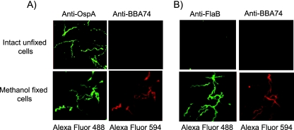
FIG. 3.
Localization of BBA74 by immunofluorescence microscopy. Intact unfixed or methanol-fixed B. burgdorferi cells were incubated with either anti-OspA and anti-BBA74 (A) or anti-FlaB and anti-BBA74 (B) antibodies. Alexa Fluor 488-labeled goat anti-rabbit IgG and Alexa Fluor 594-labeled goat anti-rat IgG were employed as secondary antibodies. Immunofluorescence staining was visualized using a Zeiss inverted Axiovert 200 microscope, and the images were acquired using AxioVision software. The images were converted to JPEG format, and brightness and contrast were linearly adjusted for each panel. The top and bottom labels indicate the primary and secondary antibodies used for each image panel. All the images were acquired at 1,000× final magnification.

