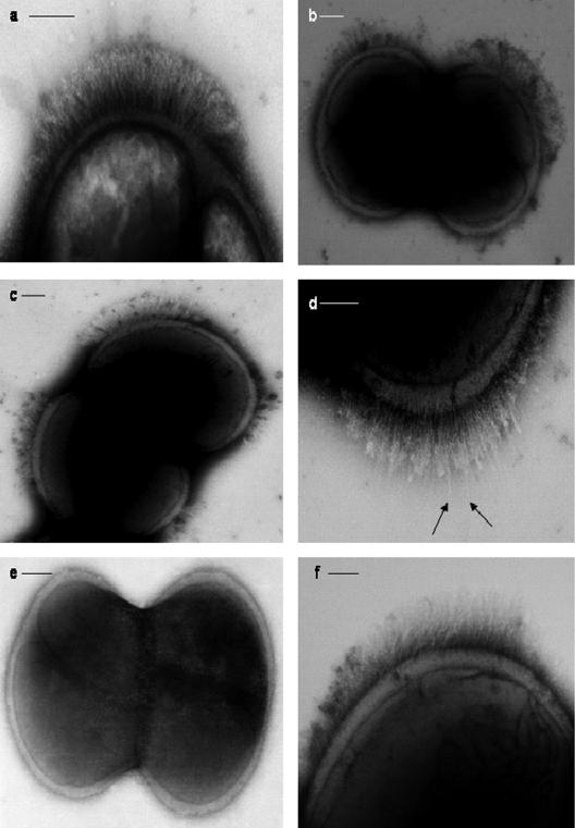
FIG. 1.
Micrographs of S. epidermidis NCTC 11047 cells (a to e) and S. epidermidis RP62A (f) negatively stained with 2% (wt/vol) methylamine tungstate. (a) One lateral tuft of fibrils on one side of the septum. No fibrils were detected on the remaining cell surface (not shown in the image). (b) Two lateral tufts of fibrils, one on either side of the septum. (c) Four lateral tufts symmetrically positioned on either side of the septum. (d) Tuft of fibrils showing a few longer individual fibrils (arrows) projecting through the mass of shorter fibrils. (e) Dividing cell without any tufts of fibrils. (f) Cell of strain RP62A carrying a tuft of fibrils. Bars, 100 nm.

