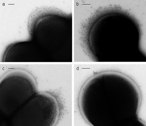
FIG. 6.
Electron micrographs of S. epidermidis NCTC 11047 cells labeled with anti-Aap antibodies and 10-nm colloidal gold-conjugated secondary antibody negatively stained with 2% (wt/vol) methylamine tungstate. (a) Fib+ cell labeled on the tuft fibrils with anti-Aap A-region antibody (top cell) attached to a Fib− cell with no tuft and no gold labeling. (b) Fib+ cell labeled with anti-Aap B-region antibody revealing a very extensive fibrillar tuft. (c) WT cells labeled with anti-A antibody, showing a labeled cell with a tuft (Fib+) attached to a cell with no labeling and no tuft (Fib−). (d) Fib− cell showing no specific gold attached after labeling with anti-A antibody. Bars, 100 nm.

