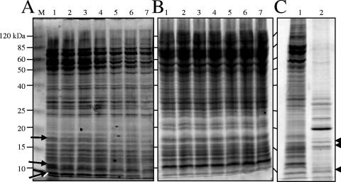
FIG. 1.
Lysis of M. xanthus cells with glass beads. Vegetative cells grown in CTTYE broth to the mid-log phase were resuspended in either TPM buffer (A) or DTT lysis buffer (B), vortexed with 0.1-mm-diameter glass beads, separated by 12% SDS-PAGE, and stained with Coomassie blue. Before protein mixtures were separated by electrophoresis, they were allowed to sit at room temperature to examine the potential of endogenous proteases to hydrolyze proteins. Lane M contained the molecular weight standard, and protein molecular masses are indicated on the left. Lane 1, zero time at room temperature; lane 2, 2 h; lane 3, 4 h; lane 4, 6 h; lane 5, 16 h; lane 6, 24 h; lane 7, 48 h. The arrows in panel A indicate protein species that were degraded over time in the TPM buffer lysate. (C) Vegetative cells (100 μg) (lane 1) and 5-day-old myxospores (10 μg) (lane 2) were lysed in DTT lysis buffer by vortexing with glass beads, separated by 12% SDS-PAGE, and stained with Coomassie blue. The arrowheads indicate protein species appearing at similar locations and in similar amounts in the two lysates.

