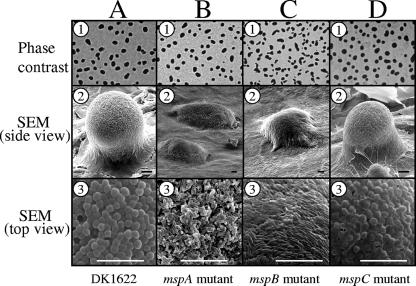
FIG. 5.
Comparison of fruiting body formation on TPM agar. Five-day-old fruiting bodies of wild-type strain DK1622 (column A) and the mspA (column B), mspB (column C), and mspC (column D) mutants are shown. Fruiting bodies were viewed by either phase-contrast microscopy (row 1) or SEM (row 2, side view; row 3, top surface of fruiting bodies). Bars = 10 μm.

