FIG. 6.
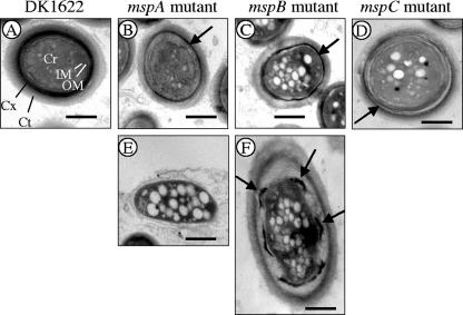
TEM comparison of ultrastructures of spores from 5-day-old fruiting bodies. Spores from fruiting bodies that had been fixed with glutaraldehyde and paraformaldehyde but had not been disrupted by sonication were examined. (A) DK1622 spore. Cr, core; IM, inner membrane; OM, outer membrane; Cx, cortex; Ct, coat. (B to D) Typical spores from mspA (B), mspB (C), and mspC (D) mutant fruiting bodies. These spores tended to have thin cortex layers (arrows). (E) Cell from the periphery of an mspA mutant fruiting body. In some spores of the mspB mutant a thin, intact cortex was observed (panel D, arrow), but in other spores the cortex appeared to be a series of disconnected patches (panel F, arrows). Bars = 500 nm.

