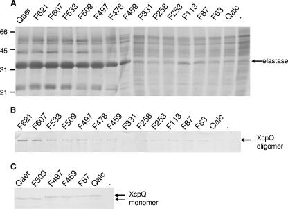
FIG. 2.
Functionality and complex formation for the different XcpQ secretins. (A) Extracellular protein profiles of P. aeruginosa strain PAN1 with constructs encoding the different XcpQ variants or with the empty vector (lane −). Secreted proteins were precipitated with 5% TCA and separated by SDS-PAGE. The position of the major protein secreted by the type II pathway, elastase, is indicated on the right, and the positions of the molecular mass standard proteins (in kDa) are indicated on the left. (B and C) Cell envelopes (B) or whole-cell preparations (C) of strain PAN1 with constructs encoding different XcpQ variants or with the empty vector (lane −) were subjected to SDS-PAGE. Proteins were transferred to nitrocellulose membranes and visualized by immunodetection with polyclonal antibodies directed against XcpQ. The positions of the XcpQ oligomers and monomers are indicated on the right.

