FIG. 4.
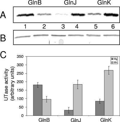
Effect on uridylylation of Mg2+ or Mn2+. The three PII paralogs (0.5 μM) were incubated with either 250 μM α-ketoglutarate and 25 mM Mg2+ (lanes 1, 3, and 5) or 60 μM α-ketoglutarate and 3 mM Mn2+ (lanes 2, 4, and 6). For both divalent cations used, 2 mM ATP, 0.13 μM GlnD, and 0.5 mM UTP, supplemented with [α-32P]UTP, were included in the reaction mixture. Samples were withdrawn from the reaction mixtures after 20 min of incubation and stopped by addition of SDS cocktail. (A) Autoradiogram showing incorporation of [α-32P]UMP into GlnB (lanes 1 and 2), GlnJ (lanes 3 and 4), or GlnK (lanes 5 and 6). (B) Coomassie-stained SDS-PAGE showing the levels of PII proteins loaded in panel A. (C) Histogram showing the difference in incorporation of [α-32P]UMP between the R. rubrum PII proteins with Mg2+ (dark) or Mn2+ (gray) in the uridylylation reaction. The amount of labeled, uridylylated GlnB, GlnJ, or GlnK was quantified using the Image Quant program. The data shown are from at least three independent experiments. UTase, uridylyltransferase.

