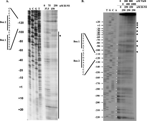
FIG. 5.
VirB displaces H-NS from the icsB regulatory sequence. (A) DNase I footprinting was used to reveal the pattern of H-NS-mediated protection of the icsB regulatory region. The vertical line shows the region of protection, and a residue showing hypersensitivity to DNase I is indicated by an arrowhead. (B) H-NS protein (250 nM) was prebound to the same icsB sequence. VirB protein was added in increasing concentrations (0 to 1,000 nM). Bases protected from DNase I cleavage are indicated by white arrowheads, and those showing hypersensitivity to DNase I in the presence of VirB are indicated by black arrowheads. In each gel, a DNA sequencing ladder generated using the same oligonucleotide primer used to generate the probe for footprinting is shown, as are the locations of the box 1 and box 2 motifs described in the legends to Fig. 1 and 2.

