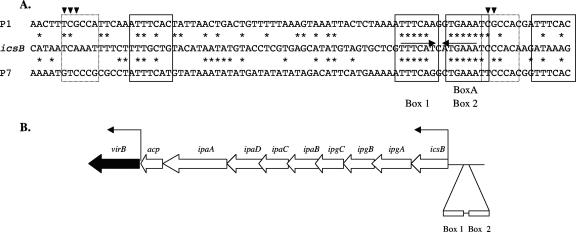
FIG. 7.
(A) Alignment of the icsB regulatory region with the parS sequences of phages/plasmids P1 and P7. The DNA sequence of the promoter-distal portion of the icsB regulatory region that contains boxes 1 and 2 is aligned with the parS sequences of phages/plasmids P1 and P7. The converging horizontal arrows show the inverted repeats associated with boxes 1 and 2. The four heptameric and two hexameric parS motifs involved in ParB protein interaction are boxed by solid- and dotted-line rectangles, respectively. Downward-pointing arrowheads indicate residues within the hexamers that allow ParB proteins to distinguish different parS sequences. The asterisks indicate residues that are conserved between the icsB regulatory region and the parS sequences. (B) Genetic map of the portion of the large virulence plasmid showing the relative locations of the virB gene and the regulatory sequences of the icsB-ipg-ipa-acp operon. The angled arrows represent promoters. The relative positions of the box 1 and box 2 motifs upstream of the icsB promoter are shown. The diagram is not drawn to scale.

