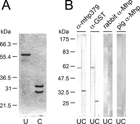
FIG. 1.
SDS-PAGE analysis and Western immunoblotting of recombinant GST-mhp379 expressed in E. coli and purified by affinity chromatography. (A) Coomassie brilliant blue-stained gel of recombinant GST-mhp379 (lane U) and thrombin-cleaved recombinant GST-mhp379 (lane C) fusion products separated by SDS-10% PAGE with molecular mass markers (Novex). (B) Western blots of uncleaved (lanes U) and thrombin-cleaved (lanes C) recombinant GST-mhp379 probed with rat antiserum raised against gel-purified thrombin-cleaved recombinant mhp379 (α-mhp379), rabbit antiserum raised against GST (α-GST), rabbit antiserum raised against M. hyopneumoniae strain LKR whole-cell proteins (rabbit α-Mhp), and antiserum from a pig infected with M. hyopneumoniae strain Adelaide Beaufort (pig α-Mhp). Proteins were separated by SDS-10% PAGE with prestained molecular mass markers (New England BioLabs) and Western transferred.

