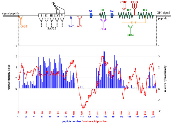
Figure 5.
Overview of PrPC secondary structures and antibody epitopes versus peptide-array binding pattern and Kyte-Doolittle hydrophilicity plot. Schematic representation of PrPC showing signal sequences, β-sheets (S1, S2), α-helices (H1, H2, H3), disulfide bridge site (S-S), glycosylation sites (CHO) and relative positions of the antibodies used in this study. The sequence of PrP is lined up with both the Kyte-Doolittle hydrophilicity plot (negative = hydrophobic and positive = hydrophilic) and the relative binding pattern found with the ovine peptide-array.

