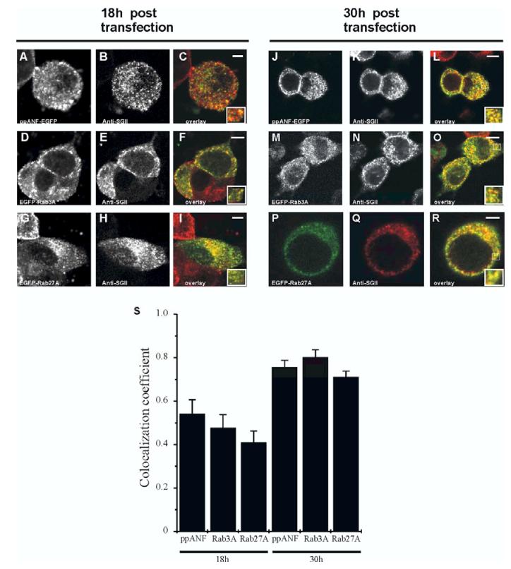
Figure 2.
Improved co-localisation between ppANF-EGFP/EGFP-Rab3A/EGFP-Rab27A and secretory granules over time post-transfection. PC12 cells were transfected with ppANF-EGFP (A-C, J-L), EGFP-Rab3A (D-F, M-O) or EGFP-Rab27A (G-I, P-R), and fixed 18 hours or 30 hours following transfection as indicated. After fixation, cells were immunostained with anti-secretogranin II (anti-SGII). Images are composites of green, EGFP fluorescence, and red, anti-SGII immunofluorescence. Areas of overlap appear in yellow and increase from 18 to 30 hours post-transfection. The scale bars represent 4 μm. S, Quantification of the colocalisation of EGFP and SGII was carried out using ImageJ and the data are shown for the correlation coefficient as mean ± SE (n=5).

