
Figure 3.
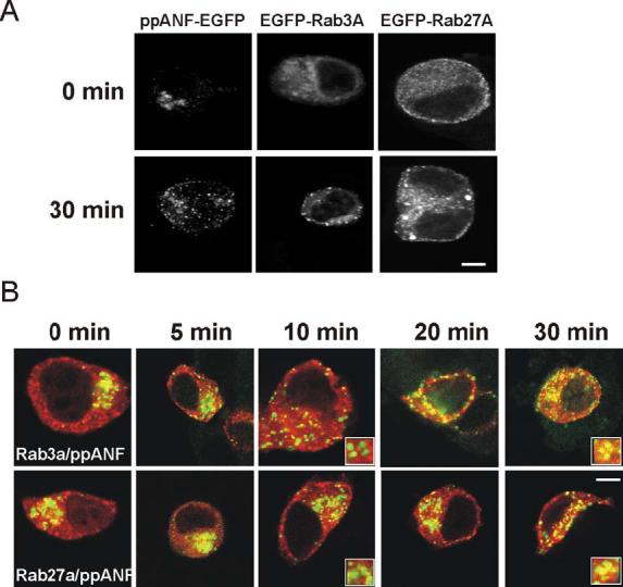
Appearance of Rab3A and Rab27A on immature secretory granules during granule biogenesis and maturation. PC12 cells were transfected with ppANF-EGFP, EGFP-Rab3A, or EGFP-Rab27A (A) and cultured for 18h at 20°C to block granule budding at the TGN and then fixed (time 0) or incubated for 30 min at 37°C to allow budding of immature granules to proceed. PC12 cells were co-transfected with ppANF-EGFP and mRFP-Eab3A or mRFP-Rab27A, cultured for 18h at 20°C and then fixed at the indicated times after incubation at 37°C. The images show overlays of ppANF-EGFP fluorescence in green mRFP fluorescence in red. Expanded inserts are shown for the 10 and 30 in time points to show lack of co-localisaton at 10 min and co-localisation at 30 min. The scale bars represent 4 μm.