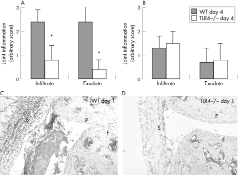
Figure 2 Frontal sections of whole knee joints 1 and 4 days after induction of immune complex‐mediated arthritis (ICA) in TLR4−/− mice and their wild‐type controls. The inflammatory cell mass present in the synovium (infiltrate) and in the knee joint cavity (exudate) was determined using an arbitrary scale from 0 to 3: 0, no cells; 1, minor; 2, moderate; 3, maximal. The inflammatory cells were counted independently by two observers blinded to the experimental set‐up. Data are the mean of six mice. Two independent experiments were performed. Significance was tested using the Wilcoxon rank test (*p<0.05). Original magnification of the photographs is ×250. F, femur; P, patella; S, synovium. Note the significantly lower inflammatory mass in knee joints of TLR4−/− mice (A and photograph D v wild‐type control A and photograph C) at day 1 after ICA induction. No differences were found at day 4 (B).

An official website of the United States government
Here's how you know
Official websites use .gov
A
.gov website belongs to an official
government organization in the United States.
Secure .gov websites use HTTPS
A lock (
) or https:// means you've safely
connected to the .gov website. Share sensitive
information only on official, secure websites.
