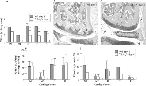
Figure 3 Measurement of cartilage destruction: proteoglycan depletion (A–C) at day 1 and matrix metalloproteinase (MMP)‐mediated cartilage destruction (expression of VDIPEN neoepitopes) (D) and chondrocyte death (E) at day 4 after ICA induction in knee joints of TLR4−/− and their wild‐type controls. Proteoglycan depletion was determined in various cartilage layers of the knee joint (P, patella; MF, medial femur; MT, medial tibia; LF, lateral femur; LT, lateral tibia) and was significantly lower in arthritic knee joints of TLR4−/− at day 1 after arthritis (A and photographs C v control B). F, femur; M, meniscus and T, tibia. VDIPEN staining was measured at day 4 and expressed as percentage positive staining of the total cartilage area. No significant differences were found between TLR4−/− and their wild‐type controls at day 4 after ICA induction. Chondrocyte death was expressed as percentage of empty lacunae of the total cartilage area. Note that chondrocyte death was lower in TLR4−/− mice, although values did not reach significance (E). Data represent the mean (SD) of seven mice and were statistically evaluated using the Wilcoxon rank test. *p<0.05.

An official website of the United States government
Here's how you know
Official websites use .gov
A
.gov website belongs to an official
government organization in the United States.
Secure .gov websites use HTTPS
A lock (
) or https:// means you've safely
connected to the .gov website. Share sensitive
information only on official, secure websites.
