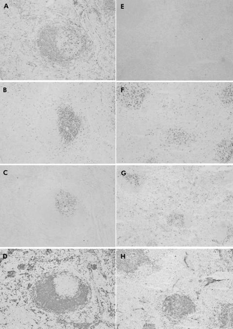
Figure 1 Immunohistochemical staining for the spleen of wild‐type (WT) (A–D) and Aα0/0 mice (E–H) using antibodies for I‐A (A, E), CD4 (B, F), CD8 (C, G), and B220 (D, H). Many I‐A positive cells (A) were observed, mainly in the B220+ site (D) in the spleen of WT mice. Despite many B220+ cells (H), no I‐A positive cells were observed in Aα0/0 mice (E). The number of CD8+ cells was similar between WT mice (C) and Aα0/0 mice (G) but the number of CD4+ cells (F) was much smaller in Aα0/0 mice than in WT mice (B). Original magnification: A–H ×100.

An official website of the United States government
Here's how you know
Official websites use .gov
A
.gov website belongs to an official
government organization in the United States.
Secure .gov websites use HTTPS
A lock (
) or https:// means you've safely
connected to the .gov website. Share sensitive
information only on official, secure websites.
