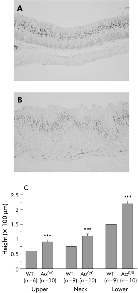
Figure 3 Proliferation status of gastric corpus mucosal cells using Ki‐67 immunohistochemistry. There was a characteristic Ki‐67 staining pattern restricted to the proliferating neck zone in the mucosa of wild‐type (WT) mice (A). In contrast, the proliferative compartment was considerably expanded in Aα0/0 mice (B). At six months old, the height of the upper, neck, and lower zones in Aα0/0 mice, as divided by Ki‐67 signals, was significantly greater than that in WT mice (p<0.001) (C). Results are expressed as mean (SEM). n = number of mice used. ∗∗∗p<0.001 compared with the same mucosal zone of six month old WT mice by the Student's t test. Original magnification: A, B ×100.

An official website of the United States government
Here's how you know
Official websites use .gov
A
.gov website belongs to an official
government organization in the United States.
Secure .gov websites use HTTPS
A lock (
) or https:// means you've safely
connected to the .gov website. Share sensitive
information only on official, secure websites.
