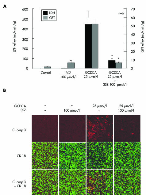
Figure 6 Sulfasalazine reduced bile acid induced liver injury and apoptosis in perfused rat livers. Rat livers were perfused with 25 µmol/l glycochenodeoxycholic acid (GCDCA) or the carrier dimethyl sulfoxide only, in the absence or presence of 100 µmol/l sulfasalazine (SSZ) for 60 minutes. (A) After 55 minutes of bile acid administration, lactate dehydrogenase (LDH) and glutamate‐pyruvate transaminase (GPT) activities in the hepatovenous effluate were determined photometrically. Results are mean (SD) of six independent experiments. (B) Hepatocyte apoptosis was determined by immunohistochemistry of activated cleaved (Cl) caspase 3 and double immunofluorescence labelling for activated caspase 3 and cytokeratin (CK) 18. After 60 minutes, double immunofluorescence labelling for CK 18 (green) and activated caspase 3 (red and yellow due to colocalisation with CK 18) in GCDCA treated livers revealed massive hepatocellular activation of caspase 3 in cells with CK intermediate filament breakdown and granular cytoplasmic condensation. In contrast, immunoreaction for activated caspase 3 was similar to controls in livers perfused with GCDCA and SSZ (magnification ×400). Representative images from six independent experiments are shown.

An official website of the United States government
Here's how you know
Official websites use .gov
A
.gov website belongs to an official
government organization in the United States.
Secure .gov websites use HTTPS
A lock (
) or https:// means you've safely
connected to the .gov website. Share sensitive
information only on official, secure websites.
