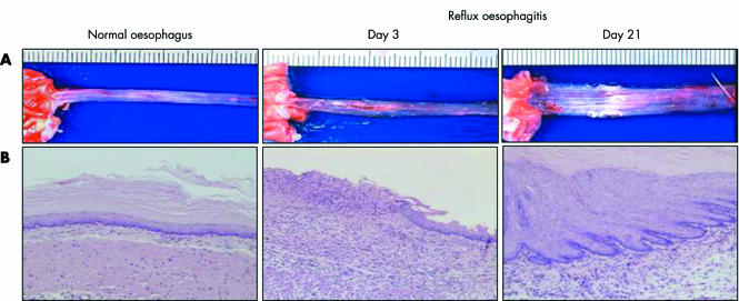
Figure 1 Macroscopic (A) and histological appearance (B) of normal oesophagus and reflux oesophagitis. Normal oesophagus has a thin epithelial layer with few inflammatory cells. However, several erosions and ulcers, and histological defects of the epithelium and marked inflammatory cell infiltration are observed on day 3 (acute phase). Oesophageal lesions with mucosal thickening with basal cell hyperplasia, elongation of lamina propria papillae, and inflammatory cell infiltration were found in the middle and lower parts of the oesophagus on day 21 (chronic phase).

An official website of the United States government
Here's how you know
Official websites use .gov
A
.gov website belongs to an official
government organization in the United States.
Secure .gov websites use HTTPS
A lock (
) or https:// means you've safely
connected to the .gov website. Share sensitive
information only on official, secure websites.
