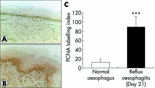
Figure 2 Oesophageal epithelial proliferation assessed by proliferating cell nuclear antigen (PCNA) staining. There were few PCNA positive cells in the normal oesophagus (A) while most epithelial cells of the basal layer and some inflammatory cells exhibited a reaction for PCNA on day 21 after induction of oesophagitis (B). The PCNA labelling index was significantly higher in oesophagitis than in normal oesophagus (C). Values are mean (SD) of six experiments.***?p <0.0001 versus normal oesophagus.

An official website of the United States government
Here's how you know
Official websites use .gov
A
.gov website belongs to an official
government organization in the United States.
Secure .gov websites use HTTPS
A lock (
) or https:// means you've safely
connected to the .gov website. Share sensitive
information only on official, secure websites.
