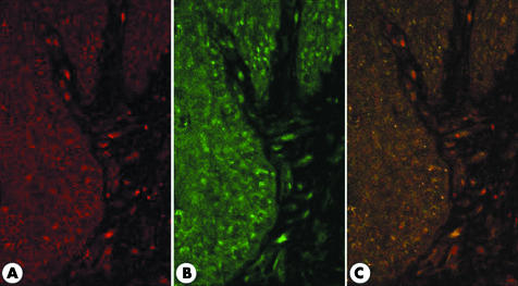
Figure 5 Confocal double immunostaining for cyclooxygenase 2 (COX‐2) and microsomal prostaglandin E synthase 1 (mPGES‐1). (A) COX‐2 (red) was stained in epithelial cells of the basal layer, and inflammatory and mesenchymal cells in the lamina propria. (B) The pattern of expression of mPGES‐1 (green) was similar to that of COX‐2. (C) Merging panels (A) and (B) revealed that COX‐2 and mPGES‐1 were colocalised.

An official website of the United States government
Here's how you know
Official websites use .gov
A
.gov website belongs to an official
government organization in the United States.
Secure .gov websites use HTTPS
A lock (
) or https:// means you've safely
connected to the .gov website. Share sensitive
information only on official, secure websites.
