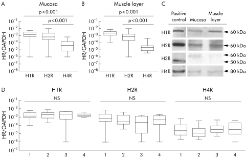
Figure 1 Histamine receptor (HR) mRNA expression in 66 human intestinal tissue samples taken from surgical specimens from 33 individuals. Data were generated by quantitative reverse transcription‐polymerase chain reaction and are displayed as glyceraldehyde‐3‐phosphate dehydrogenase (GAPDH)/HR ratio (p values as indicated). (A, B) HR expression in the mucosa/submucosa (A) and the muscular layer (B). H3R was not detected in >90% of samples. (C) Protein expression of HR in intestinal tissue homogenates, shown by immunoblot. Mucosa and muscular layer samples of colonic tissue were probed with H1R, H2R, and H4R antibodies. Human umbilical vein endothelial cells, gastric mucosa homogenate, human neuroblastoma cells MHH‐NB 11, and purified human intestinal mast cells were used as positive controls for H1R, H2R, H3R, and H4R, respectively. As indicated, H3R antibodies detected an unspecific protein band of approximately 60 kDa in lysates of human colon while a specific band migrated at the expected weight of 50 kDa. (D) Comparison of HR expression at different intestinal sites: 1 = duodenum, 2 = colon, 3 = sigmoid, 4 = rectum.

An official website of the United States government
Here's how you know
Official websites use .gov
A
.gov website belongs to an official
government organization in the United States.
Secure .gov websites use HTTPS
A lock (
) or https:// means you've safely
connected to the .gov website. Share sensitive
information only on official, secure websites.
