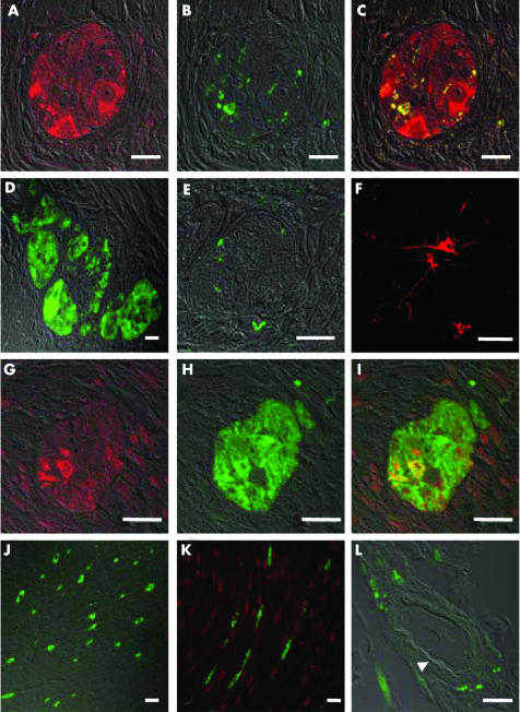
Figure 4 Immunofluorescence staining of human intestinal tissue and human brain cortex, examined by confocal microscopy. (A–C) Myenteric ganglion cells in human colon stained with PGP 9.5 (A, red), tyrosine hydroxylase (B, green), or both (C—(A) and (B) merged). (D) PGP 9.5 (green) and histamine 3 receptor (H3R) (red) double staining of a myenteric ganglion in human colon; no H3R staining was observed. (E) Tyrosine hydroxylase (green) and H3R (red) double staining of a myenteric ganglion in human colon; sympathetic nerve endings did not express H3R. (F) H3R staining (red) of neurones in human brain cortex. (G–I) Myenteric ganglion cells in human colon stained with anti‐H1R antibody (G, red), PGP 9.5 (H, green), or both (I—(G) and (H) merged). Note the prominent co‐localisation of H1R and PGP 9.5 staining. (J) PGP 9.5 (green) and H3R (red) staining of the circular muscle layer; nerve fibres (green) are devoid of H3R. (K) PGP 9.5 (green) and H1R (red) staining of the circular muscle layer; H1R was expressed on smooth muscle but not on nerve fibres. (L) PGP 9.5 (green) and H3R (red) staining of a submucosal blood vessel; after subtraction of unspecific autofluorescence there remained a faint red staining of a circular structure (arrowhead), reminiscent of the basal lamina.

An official website of the United States government
Here's how you know
Official websites use .gov
A
.gov website belongs to an official
government organization in the United States.
Secure .gov websites use HTTPS
A lock (
) or https:// means you've safely
connected to the .gov website. Share sensitive
information only on official, secure websites.
